The human brain is the most remarkable organ on planet earth. It’s the defining feature of the human species.
How did the human brain come to be?
The first brain appeared in the fossil record 550 million years ago. Scientists don’t truly understand the exact way that brains evolve. Across different species, brain parts are used for certain functions and basic behaviors. For example, mice, chickens, and apes all have different brain structure due to the function require.
The parts of the human brain are unique from anything else. From the time of birth, the newborn brain begins to direct deep survival functions. These include breathing, heart rate, muscle movement, and the senses.
Higher brain function includes feeding, hunger, digestion, hormones, and sleep cycles. In higher consciousness mammals, it projects emotions, feelings, communications, creativity, and memories.
Today research is beginning to unearth the vast complexity of the human brain.
A glance at the function of the human brain parts
- At 2% of our body weight, humans have the largest brain of all vertebrates relative to body size.
- The human brain weighs about 3.3 lbs. (1.5 kilograms)
- The human brain contains close to 86 billion nerve cells (neurons) — the “grey matter.”
- The human brain has billions of nerve fibers (axons and dendrites) — the “white matter.”
- These neurons are connected by trillions of connections or synapses.
A summary of the function of brain parts
As a general rule, the function of brain parts arrange from simple to complex. These are located from inside of the brain to the brain surface.
The outer parts control the more emotional and thought based functions. While the inner and lower parts of the brain control simpler and more basic functions like breathing and blood pressure.
Now we will take a trip through the different human brain parts, exploring their function.
The Cerebrum
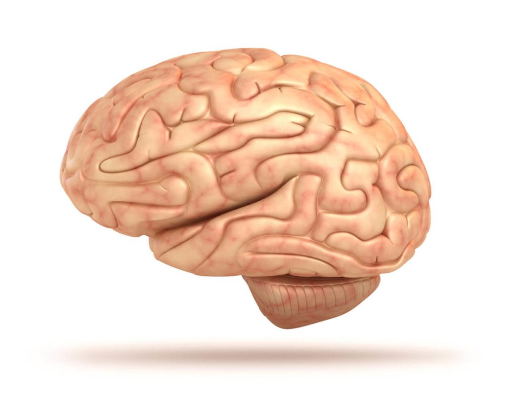
The Cerebrum is the largest human brain part and makes up around 80% of its total volume.
The cerebrum is the outer ‘folded’ section of the brain. It is what you would recognize by its walnut like appearance.
It’s the largest brain part in humans and accounts for about 80% of the brain’s mass. It is divided into two sides, the left and right hemispheres. The hemispheres are separated by a deep groove down the center from the back of the brain to the forehead.
These two halves are connected by long neuron branches called the corpus callosum. It is relatively larger in women’s brains than in men’s. The cerebrum is positioned over and around most other brain parts and structures. It has four lobes that have different functions but are also richly connected.
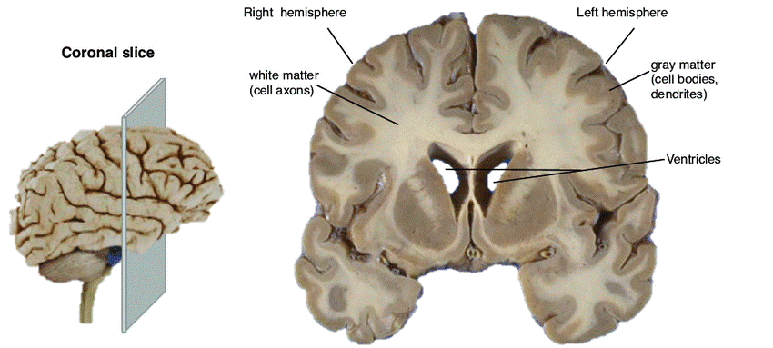
A Coronal slice of a human brain.
The outer 3 millimeters of “gray matter” is called the cerebral cortex. Here consists of tightly packed neurons that control most of our body functions. These include the mysterious state of consciousness, the senses, the body’s motor skills, reasoning, and language.
The Cerebral Cortex
The cerebral cortex structure is divided into four sections.
These are known as lobes:
- Frontal lobe
- Parietal lobe,
- Occipital lobe
- Temporal lobe
Each associated with different functions ranging from reasoning to auditory perception.
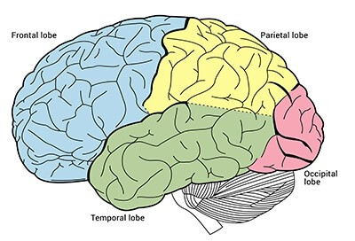
Brain structure of the cerebral cortex is divided into four lobes with different functions.
The cerebral cortex as a brain part in humans is a recent development in the course of evolution. It handles the many signals responsible for perception, movement, and mental processes.
Let’s look at the structure and function of each of the brain lobes.
The Frontal Lobe
The part of the brain located at the front of the head or forehead. It functions across the control of motor skills, higher level cognition, and expressive language.
The frontal lobe is the part of the brain that most manages ‘human functions.’ It’s most complex and recently evolved part of the brain. Out of all the cerebral cortex, it also develops last in young adulthood.
The two main parts of the frontal lobe are:
- Prefrontal cortex (PFC) is the cerebral cortex which is the front part of the frontal lobe. This brain region runs planning complex cognitive behavior, personality, decision making, and moderating social behavior.
- Motor Cortex – At the back of the frontal lobe, near the central sulcus, lies the motor cortex. This area of the brain receives information from various lobes of the brain. It combines all of this information to carry out body movements.
Other divisions of the frontal cortex are:
- Orbitofrontal cortex (OFC): is the area of the prefrontal cortex that sits just above the orbits (or eye sockets). It is thus found at the very front of the brain and manages emotional impulses in socially appropriate ways. It creates productive behaviors including emotions, empathy, altruism, understanding facial expressions.
- Dorso-lateral prefrontal circuit (dIPFC): is the brain’s top executive. It’s a region in the frontal lobes toward the top and side: hence dorso (top) and lateral (side). It organizes responses to complex problems. These include planning the steps of an objective and searching memory for relevant experience. Adapting strategy for new data and guiding behavior with verbal skills. All of these happen while housing working memory.
Damage via stroke to the frontal lobe can lead to changes in sexual habits, socialization, and attention as well as increased risk-taking.
The Temporal Lobe
Located on the bottom section of the brain closest to the ear. This lobe is also the location of the primary auditory cortex. It is important for interpreting sounds and language. It crosses both hemispheres of the brain, helps process sensory input, including pain and sound stimuli.
It controls memory storage, emotion, hearing, and, on the left side, language.
The hippocampus is also located in the temporal lobe. It’s a part of the brain that is also heavily associated with the formation of memories.
Damage to the temporal lobe can lead to problems with memory, speech perception, and language skills.
The Parietal Lobe
Located at the upper back part of the brain. It processes sensory information to do with taste, temperature, pressure, touch, and pain.
The parietal lobe is where information is integrated or processed. It contains the somatosensory cortex, essential to the processing of the body’s senses.
Humans would not be able to feel sensations of touch if the parietal lobe was damaged.
Two functions of the parietal lobe include:
- Combines sensory information to form a single perception (cognition).
- Constructs a spatial coordinate system to represent the world around us. Individuals with damage to the parietal lobes often show striking deficits. These include problems with body image and spatial relations.
Damage to the left parietal lobe can result in what is called “Gerstmann’s Syndrome.” It includes right-left confusion.
The Occipital Lobe
Located at the back portion of the brain above the neck. It’s associated with interpreting visual stimuli and information. As home to the primary visual cortex, it receives and interprets data from the retinas of the eyes. It routes the visual data to other parts of the brain for identification and storage.
Damage to this lobe can cause visual problems such as difficulty recognizing objects, an inability to identify colors, and trouble recognizing words.
The Brain Stem
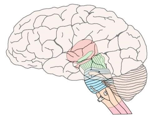
The part of the brain that connects to the spinal cord. The brain stem controls basic function of the survival for all animals. These include heart rate, breathing, digesting foods, and sleeping.
It appears earliest regarding evolution and hence controls the critical and basic functions of the human brain.
Parts and function of the Brain Stem
1) The Thalamus
Located at the top of the brain stem. The thalamus acts as a two-way relay station. It sorts, processes, and directs signals from the spinal cord and midbrain structures up to the cerebrum. Then it conveys messages from the cerebrum down the spinal cord to the nervous system.
The thalamus processes and transmits movement and sensory information. It also takes sensory information and passes it on to the cerebral cortex. The cerebral cortex also sends data to the thalamus, which then sends this information to other systems.
2) The Midbrain
Often considered the smallest brain part. It acts as a sort of relay station for auditory and visual information. The midbrain controls many vital functions such as the visual and auditory systems as well as eye movement.
Parts of the midbrain include the red nucleus and the substantia nigra. Their function is to control body movement. The darkly pigmented substantia nigra contains a large number of dopamine-producing neurons. It’s the main neurotransmitter that helps movement function.
3) The Hindbrain
The area most neuroscientists call the proto-reptilian brain. The purpose of this brain part is to coordinate basic homeostatic functions. The pons and medulla are major structures found there.
4) The Pons
The connection between the medulla and midbrain. The pons functions as a message station between several areas of the brain. It helps relay messages from the cortex and the cerebellum. Without the pons, the brain can’t function properly as messages would not be able to be transmitted.
The pons plays a key function in sleep and dreaming. The deep sleep state (REM sleep) where dreaming is most likely to occur, starts in the pons.
Injury to the pons may result in sleep disorders, sensory problems, arousal dysfunction, and coma.
Damage to the pons can also cause difficulty with sense of balance, walking, sense of touch, swallowing and speaking.
5) The medulla or medulla oblongata
Located directly above the spinal cord in the lower part of the brain stem. It controls many vital autonomic functions such as heart rate, breathing, and blood pressure.
Functions of the medulla are performed without thought. We would not be able to live without the medulla because the critical tasks it performs. These include regulating blood pressure and breathing.
6) Hypothalamus
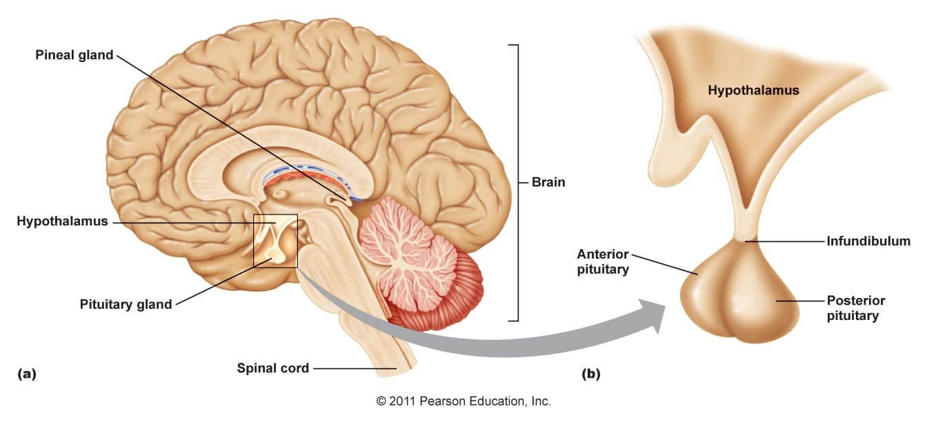
The hypothalamus acts as a link to the pituitary gland.
Located under the thalamus. The hypothalamus is a group of nuclei that lie along the base of the brain near the pituitary gland. It’s where the brain signals and the body’s hormonal system interact.
It acts as a meeting point between the nervous and endocrine system. The function varies between emotion and physical feeling. In essence, the hypothalamus maintains the body’s status quo. It monitors many bodily functions such as blood pressure, body temperature, body weight, appetite, and thirst.
The hypothalamus connects with many other regions of the brain. It is also in control of emotions, and circadian rhythms. As a close partner to the pituitary gland, it sends messages to secrete hormones. The connection gives the hypothalamus a great deal of control over many body functions.
7) The Pituitary Gland
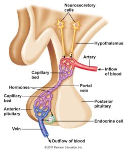
The pituitary gland connects to the base of the hypothalamus. Source
At about the size of a pea, the pituitary is the ‘master gland’ of the body. It sits in a bony hollow at the base of the brain. Just behind the bridge of your nose, it attaches to the base of your brain by a thin stalk.
It has two lobes:
a) Anterior lobe of the pituitary gland
Made up of several different types of cells that produce and release different kinds of hormones, including:
- Growth hormone
- Thyroid-stimulating hormone
- Adrenocorticotropic hormone
- Follicle-stimulating hormone
Follicle-stimulating hormone and luteinizing hormone promote the development of eggs and sperm. They hence regulate the timing of ovulation.
The anterior pituitary gland also produces several hormones with more general effects. These include human growth hormone, melanocyte-stimulating hormone (which plays a role in the pigmentation of skin), and dopamine, which inhibits the release of prolactin but is better known as a neurotransmitter.
b) Posterior lobe of the pituitary
Stores hormones that are usually produced in the hypothalamus until they’re released.
These include:
- Vasopressin: signals body to conserve water and prevent dehydration.
- Oxytocin: which stimulates the release of breast milk. It also stimulates contractions of the uterus during labor.
8) The Pineal Gland
A small, pea-shaped gland of the brain. It’s located centrally in between the two hemispheres. Its function isn’t fully understood. Researchers do know that it produces and regulates some hormones associated with sleep.
It is mainly made up of pinealocytes. These are cells that produce the hormone melatonin, which controls circadian rhythms. The secretion of melatonin varies significantly over a 24-hour cycle, from low levels during the day to a peak at night.
The pineal gland has been called the “third eye,” it contains cells very similar to the cells of the eye. It is controlled by neurons sensitive to light, which originate in the retina of each eye and end in the hypothalamus.
It responds to shifts in hours of daylight and the hormonal responses of the hypothalamus. It may also guide reproductive functions. In humans, who can conceive and give birth throughout the year, the reproductive role of the pineal gland is not fully understood. There is evidence that melatonin has a role in regulating ovulation.
Researchers found evidence that melatonin produced by the pineal gland can have a positive impact on your heart and blood pressure.
There’s some evidence that light exposure and melatonin levels may influence the menstrual cycle. Reduced amounts of melatonin may also play a role in the development of irregular menstrual cycles.
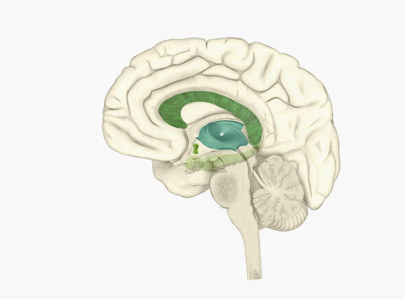
9) The Basal Ganglia
A group of large nuclei that partially surround the thalamus under the cortex (subcortical). These nuclei are important in the control of movement. The red nucleus and substantia nigra of the midbrain have connections with the basal ganglia and cerebellum which serve many important functions. These include playing a role in several autonomic functions.
Primarily used for motor control, they also are involved in motor learning, executive functions and behaviors, and emotions.
Damage to the basal ganglia network forms the basis for several movement disorders.
10) The Cerebellum
The “little brain” or two peach-size mounds of folded tissue located at the top, back of the brain stem.
It’s made up of small lobes and receives information from the balance system of the inner ear, sensory nerves, and the auditory and visual systems. It is involved in the coordination of movements as well as motor learning.
For example, the cerebellum helps control posture, balance, and the coordination of voluntary movements. It allows different muscle groups in the body to act together and produce coordinated, fluid movement. It also plays a major role in certain cognitive functions including speech.
It controls complex movements (e.g., a tennis serve). It’s also involved in some learning pathways.
The cerebellum makes up approximately 10 percent of the brain’s total size. It accounts for more than 50 percent of the total number of neurons located in the entire brain. While it controls movements, motor commands do not originate here. Instead, the cerebellum serves to modify these signals and make motor movements accurate and useful.
Stroke that restricts blood to the cerebellum include symptoms such as vertigo, headache, vomiting, and ataxia (lack of control of voluntary movement).
11) The limbic system or emotional center
The list of structures that make up the limbic system are not agreed upon.
Four of the main regions of the limbic systems include:
- The amygdala
- The hippocampus
- Regions of the limbic cortex
- The septal area
These structures relay between the limbic system and the hypothalamus, thalamus, and cerebral cortex. The hippocampus is important in memory and learning. While the limbic system itself is central in the control of emotional responses.
12) The Amygdala
Deep in the center of the limbic-emotional brain lies the amygdala. It’s the size and shape of an almond and stays constantly alert to the needs of basic survival. These include sex and emotional reactions such as anger and fear.
The amygdala inspires emotion-based response such as sweaty palms. It is larger in male brains, often enlarged in the brains of sociopaths and it shrinks in the elderly.
Recently, it’s been associated with a range of mental conditions including depression to even autism.
13) The Ventricles
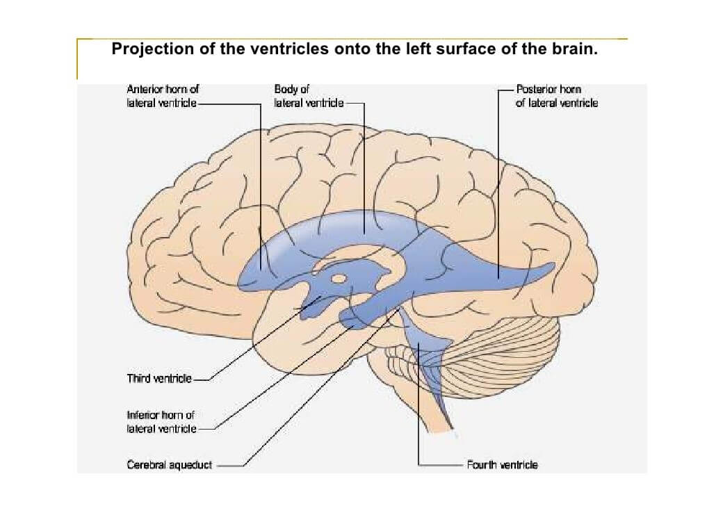
The ventricles are the part of the brain with the function to produce, carry and distribute cerebrospinal fluid (CSF).
The piping of the brain. The ventricles are a communicating network of cavities filled with cerebrospinal fluid (CSF). The ventricular system is composed of two lateral ventricles, the third ventricle, the cerebral aqueduct, and the fourth ventricle cord.
A cavity in the forebrain becomes the third ventricle, which leads further forward into the two lateral ventricles, one in each cerebral hemisphere.
Within each ventricle is a region of choroid plexus. It’s a network of ependymal cells that help to produce cerebrospinal fluid. It is continuous with the central canal of the spinal cord.
Cerebrospinal fluid is the life-source of the brain. It absorbs physical shocks to the head and neck. Distributes nutritive materials to and removes wastes from nervous tissue. It is most significant during sleep when the blood-brain barrier opens up and allows CSF to flow within the neurons.
Now it’s up to you. Do you have a good handle on the function of different brain parts?
Leave your thoughts in the comment section below.
Further reading
- https://www.ncbi.nlm.nih.gov/books/NBK234157
- https://www.researchgate.net/publication/294430587_Advanced_forward_models_for_EEG_source_imaging
- http://www.nature.com/articles/s41467-018-03464-w