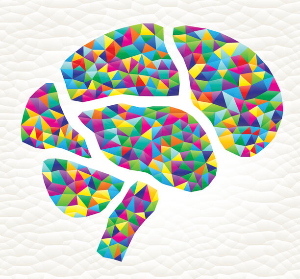
 Your brain relays between 18 and 640 trillion signals every second. The mind blowing amount of data is sent and decoded in the functional areas of the brain.
The classical idea of the human brain is the command center of your nervous system. However, research is revealing that it is far more complex than just a switchboard between your ears.
Your brain is so powerful and so diverse, it’s a big task to name all its different functions. Neuroscientists have attempted to break down the different brain parts to explain its structure.
These are known as the functional areas of the brain.
In this article, we’ll explore these different areas of the brain and their function.
How does the brain work?
Human brain structure is a hierarchy made up of about 85 billion neurons. Those neurons send signals across 18-32 trillion tiny spaces called synapses. Each synapse can convey a signal at 0.1–2 times per second. That equates to between 18 and 640 trillion signals that your brain sends and receives every second.
On top of all this, it stores some of the information it transmits as memories. As a data-storage system, the long-term hard drive of the brain may be infinite. For example, electrical impulses can trigger memories that the subject assumed to be long forgotten. It is believed that the brain can remember everything from birth through to death.
What is the main part of the brain?
The cerebral cortex is what sets apart human brains from other organisms. It’s organized at many physical scales starting at the level of single neurons and expanding up to functional systems.
Current functional imaging studies classify brain structure to three levels. These are areas, networks, and systems.
It is mostly known that the brain is organized into functional areas that are integrated into networks. However, the absolute contribution of each area to behavior is yet to be properly defined.
New large studies are integrating brain imaging and behavioral data. It allows researchers to explore broader data on brain scan patterns by comparing neural markers and functional outcomes.
Left side brain vs. right side brain
Firstly, the human brain is divided into left and right hemispheres. These are connected by a bundle of nerve fibers called the corpus callosum. The two sides are strongly though not entirely symmetrical.
The left brain controls muscles on the right-hand side of the body. Then the right brain controls the left side. One hemisphere may be slightly dominant. Like left or right-handedness.
What is the difference between left and right brain thinking?
The idea of “left brain” and “right brain” are qualities that have not been confirmed by research. There, however, some important differences between these areas. The left brain contains regions involved in speech and language (Broca’s area and Wernicke’s area). It also performs mathematical calculation and fact retrieval. It’s considered the logical or academic side of the brain.
The right brain plays a role in visual, auditory processing, and spatial skills. These generally are thought to equate to artistic ability considered more instinctive or creative in nature. However, these functions involve both hemispheres.
Image scanning shows that people use different proportions of their brain. When you use your brain you’re using both the left and right functional areas.
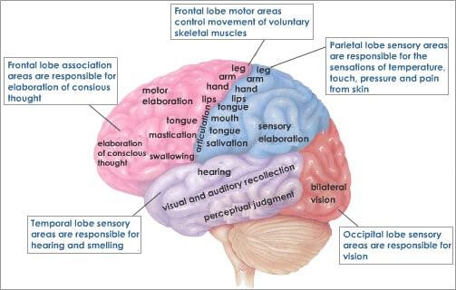
The functional areas of the cerebral cortex
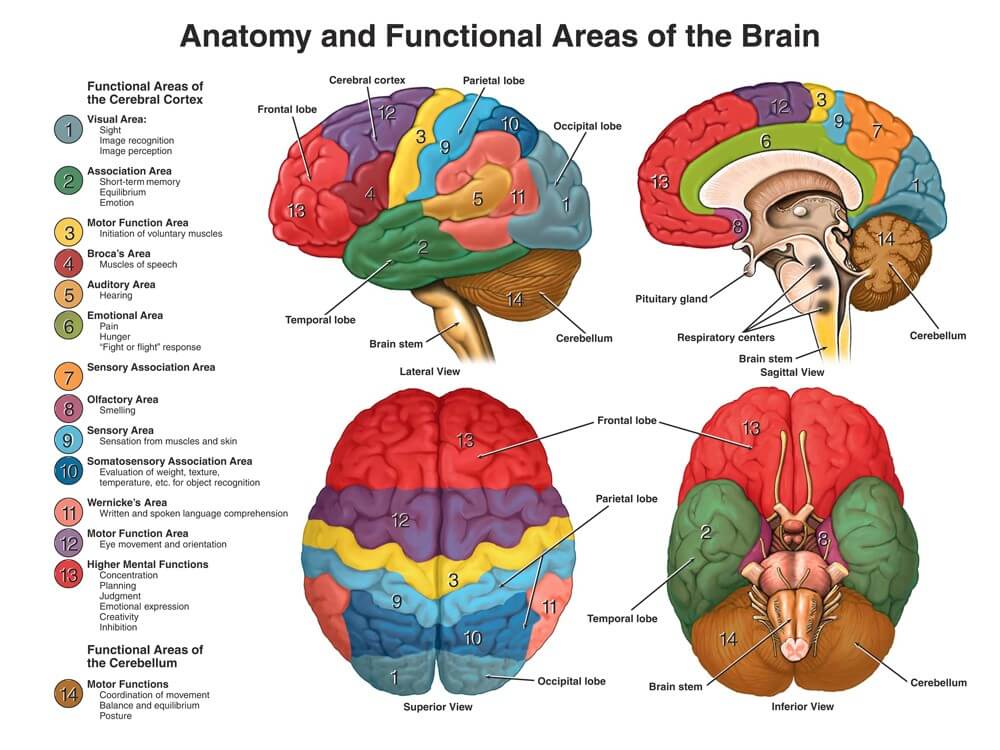
Different areas of the cerebral cortex control different functions.
The largest area of the human brain is the cerebrum. It’s also known as the cerebral cortex and controls many of the thoughts and processes we equate to the human mind. It sets us most apart from other creatures.
1) The Visual Cortex
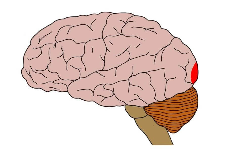
The primary visual cortex is found at the back of the human brain. This is called the occipital lobe and is located in the left and right hemispheres.
It integrates and processes visual data relayed from the retinas.
It’s divided into five different areas (V1 to V6) based on function and structure.
V1 respond to specific types of visual cues such as the orientation of edges and lines.
V2 receives and processes data from V1. Cells in this region respond to color, spatial frequency, moderately complex patterns, and orientation. V2 sends feedback connections to V1 and has feedforward connections with V3-V5.
V3 is located immediately in front of V2. Some researchers think this area is subdivided into two or three sections. Dorsal V3 receives inputs from V2 and from the primary visual area and projecting to the posterior parietal cortex. Ventral V3 (VP), has primary visual area links, and stronger connections with the inferior temporal cortex.
V4 is a lesser understood area involved with selective attention. Like V2, V4 is tuned for orientation, spatial frequency, and color. Unlike V2, V4 is tuned certain details of complexity, like simple shapes.
V5 thought to play a major role in the perception of motion, the integration of local motion signals into global percepts, and the guidance of some eye movements.
V6 appears to respond to visual stimuli associated with self-motion [and wide-field stimulation
2) Association area
Part of the temporal lobe of the cerebral cortex. It receives inputs from multiple areas; association areas integrate incoming sensory information, and also form connections between sensory and motor areas.
3) Motor function area
Located at the back of the frontal cortex (top of the head). The motor cortex sends muscle signals down the spinal cord. The left motor cortex controls the right side of the body, and right motor cortex controls the left side of the body.
It is further divided into:
- Premotor cortex – chooses the muscle group required for an action. Eg, to kick a soccer ball, it the right leg muscle group must be used.
- Primary motor cortex – sends signals the motor neurons of the spinal cord to stimulate the intended muscles.
- Supplementary motor cortex – controls movements requiring two muscle groups. Eg. Using two hands to pick up a box.
However, some of the other motor areas in the brain also play a role in certain motor functions
4) Broca’s area
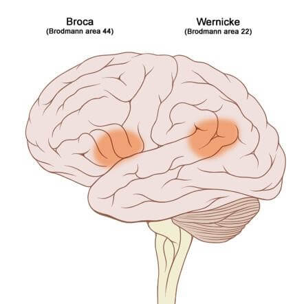
Broca’s and Wernicke’s area of the brain. Source
Located within the frontal lobe and is critical for speech production. It is larger in the left hemisphere than in the right. This size difference has been correlated with language dominance.
5) Auditory area
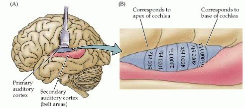
Primary Auditory area. Source
The auditory cortex receives input from the ears via lower parts of the auditory system. It then transmits signals back to these areas and other parts of the cerebral cortex.
It was subdivided into primary (A1) and secondary (A2), as well as and further association areas.
Newer divisions of the auditory cortex are:
- the core (which includes A1),
- the belt
- the para belt.
The belt is the area immediately surrounding the core. The para belt is adjacent to the lateral side of the belt
6) Emotional area
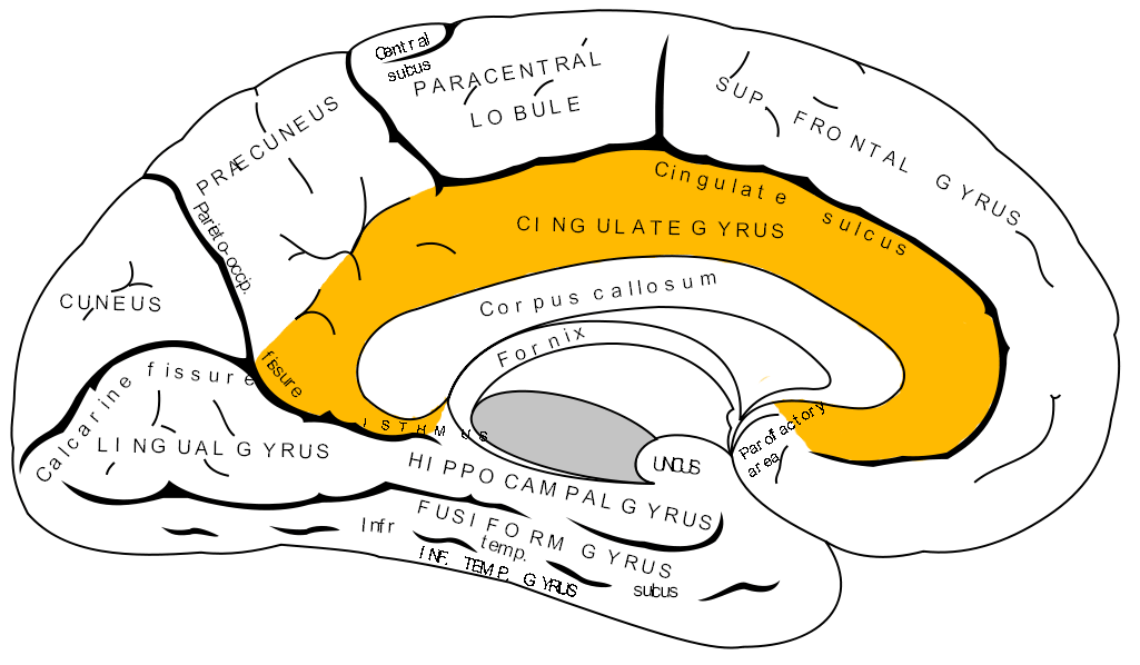
The emotional cortex links the cerebrum to the limbic system. Source
The emotional cortex is on the central underside of the cerebrum. The cingulate gyrus is the part of the cerebrum that lies closest to the limbic system, just above the corpus callosum. It conveys with the limbic system and works is the subcortical structures meet the cerebral cortex. Structures of the limbic system are involved in motivation, emotion, learning, and memory.
The limbic system includes:
- hypothalamus
- hippocampus,
- and amygdala
Known as emotional thinking, these areas are involved with what makes you laugh and problem-solving.
7) Sensory association area
An association area around the borders of primary receiving areas, where sensory stimuli are interpreted relayed.
8) Olfactory area
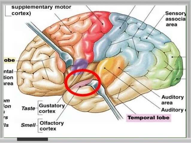
The Olfactory cortex responsible for smell function.
The general area of the brain dealing with the sense of smell. It includes the olfactory bulb, tract, and trigone, the anterior portion of the gyrus cinguli, and the uncus. Receives data directly from the nose through the anterior perforated substance.
9) Sensory Cortex
The sensory cortex, located in the front portion of the parietal lobe. It receives information relayed from the spinal cord regarding the position of various body parts and how they are moving.
This middle area of the brain can also be used to relay information from the sense of touch, including pain or pressure which is affecting different portions of the body.
The sensory cortex projects information from certain body parts to the brain.
Somatosensory cortex is located in the postcentral gyrus. It is the main sensory receptive area for the sense of touch. A map of sensory space divided for sensory areas in this location is called the sensory homunculus.
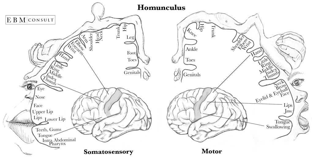
The somatosensory homunculus maps out sensory input and output to parts of the human body.
10) Wernwicke’s area
The region of the brain that is important for language development. It is located in the temporal lobe on the left side of the brain and processes the understanding of speech. It works with Broca’s area which formulates speech.
11) Motor function area (eye movement and coordination)
The area of the frontal cortex in animals over which stimulates eye movements.
12) Frontal lobe – Higher and Human Function
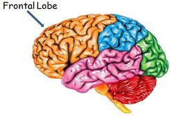
The frontal lobe is where higher human-like brain activity occurs.
Where higher mental processes such as thinking, decision making, and planning. Highly developed in humans, the frontal lobe is also where our personality is formed and where we can carry out higher mental processes such as planning.
In addition, the frontal lobe is necessary to be able to speak fluently (without fault) and meaningfully.
13) Cerebellum motor functions
Responsible for balance and coordination of muscles and the body. Critical for being able to perform everyday voluntary (done with purpose and intent) tasks such as walking and writing.
Further reading:
- https://www.ncbi.nlm.nih.gov/books/NBK482504/
- https://www.ncbi.nlm.nih.gov/pmc/articles/PMC2705206/
- https://www.sciencedirect.com/science/article/pii/S1364661318300238